CHEST 50 by Dr Sushen Kumar, Nanavati Superspeciality Hospital, Mumbai
4 year old male with progressive shortness of breath.
History of hospitalization for pulmonary adenovirus infection at 10 months of age
Select option
A - Bronchiolitis obliterans
B - Congenital Lobar Emphysema
C - Dependent opacities
D - Broncho alveolar Carcinoma
click for answer
Findings:
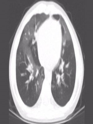
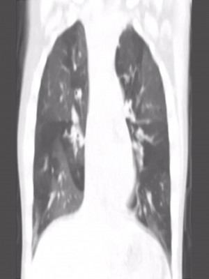
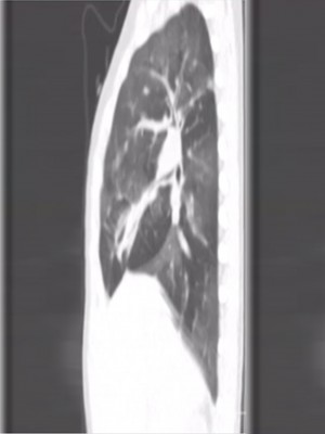
Axial, coronal and sagittal CT images show sharply defined areas of mosaic attenuation with decreased calibre of the vessels in the hyperlucent lobes or segments.
These changes represent combination of air trapping and oligemia.
Diagnosis: Bronchiolitis obliterans
Discussion:
Bronchiolar and peribronchiolar inflammation of the bronchioles leads to submucosal and peribronchiolar fibrosis. This causes an obstruction of the bronchial lumen, also known as obliterative bronchiolitis. Associated with transplantation (Eg: bone marrow/ graft vs host-uptp 10%; lung transplant), viral infection(Known as Swyer-James syndrome or MacLeod syndrome in children), toxin inhalation, rheumatoid arthritis, inflammatory bowel disease and drug reactions (bleomycin, gold, cyclophosphamide, methotrexate and amiodarone).
Airtrapping on expiratory scan( ie. Mosaic perfusion pattern)- this is classic and due to obstruction of the small airways. Bronchiectasis (Ie.dilated and thick walled).Centrilobular groundglass opacification. Expiratory HRCT differentiates between the two causes(ie. Small airway disease vs pulmonary vascular abnormalities)