15 Aug, 2018
Value of MRI in characterizing adnexal masses
Introduction
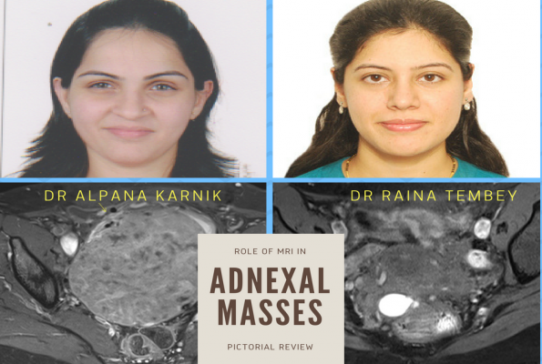
Adnexal masses, both incidental and symptomatic pose a challenging diagnostic problem because many times, the imaging features on ultrasound may overlap. Thus, once an adnexal lesion has been detected, the goal of further imaging is accurate tissue characterization resulting in surgery only for lesions that are indeterminate or frankly malignant [1]. Magnetic resonance imaging (MRI) and even Computed tomography (CT) play an increasingly important role in characterization of adnexal lesions, especially when they are large and indeterminate on ultrasound. This pictorial essay discusses the value of characterization of adnexal masses by MRI.
Patient Preparation & Technique
The patient does not necessarily have to be fasting for the study. A baseline Serum Creatinine report is requested, in case there may be a need for intravenous contrast (Gadolinium -Gd). The superior tissue contrast and flexible imaging planes afforded by MRI versus competing technologies permit optimal depiction of the pelvic viscera [2] Basic imaging protocol must include a high-resolution, free-breathing (FSE) T2 weighted (T2WI) sequences in the axial, sagittal, and coronal planes, axial T1 weighted imaging (T1WI) sequence with and without fat suppression, and diffusion-weighted imaging (DWI) to characterize complex ovarian lesions, and small metastatic lymph node and peritoneal deposits in the pelvis [3,4].
Normal Anatomy
The uterus is seen as a homogenous, medium-signal intensity structure, with the myometrium and endometrium separated by a junctional zone that is in the inner third of the myometrium, and separates the endometrium and outer myometrium [3]. The ovaries are isointense on T2WI and are demarcated by the presence of follicles in the reproductive age group, that are seen as hyperintense on T2WI(Figure 1).
The fallopian tubes unless enlarged or diseased are not distinctly visualized on MRI sequences.
Ovarian Cysts
While simple ovarian cysts are imaged almost completely by ultrasound, whether in the reproductive age group or post menopausal women, the presence of a cyst greater than 7 cm needs further imaging, either as a prelude to intervention, or for better characterization [5].
MRI usually shows hypointense T1WI signal with hyperintense T2WI signal, suggestive of clear fluid. Serous cystadenomas have similar imaging characteristics, while mucinous cystadenomas present as multilocular (honeycomb like locules) lesions with a thin regular wall with septae.
Hemorrhagic corpus luteum cysts have a characteristic appearance of blood products with hyperintense signal on T1WI and iso to hyperintense signal on T2WI [3].
Para ovarian cysts can also be easily differentiated by visualizing the normal ovary separately next to the cyst. (Figure 2).
.jpg)
Pedunculated or Subserous Uterine fibroids
Pedunculated uterine subserosal and broad-ligament fibroids frequently present as adnexal masses and ultrasound is usually adequate. However, in indeterminate cases, MRI helps in the diagnosis of these lesions by showing their extraovarian location and their connection to the uterus or the broad ligament.Fibroids can undergo various types of degeneration, such as cystic, hyaline, mucinous, myxomatous, fatty and carneous (red), resulting in a wide range of observed MRI signal intensities [1,6 ](Figure 3).
.jpg)
The common MRI features of fibroids are that they are round, well-demarcated, displace rather than infiltrate surrounding structures, and often show homogenous signal intensity and pattern of enhancement [6].
Endometriosis
MRI is superior to ultrasound in identifying sites of disease hidden by dense adhesions and this has made pelvic MRI the non-invasive imaging technique of choice in endometriosis. T1WI fat-suppressed sequence increases the detection of small implants by allowing better definition, as well as differentiation between hemorrhagic and fat components (dermoid) [1,3,7]. Large cysts containing hemorrhage can be easily noted on MRI as hyperintenseon T1WI with no signal change on fat suppressed images (Figure 4).
.jpg)
.jpg) Involvement of the adjacent uterine ligaments with endometriotic nodules, adhesions, and pelvic deposits in the pouch of Douglas (POD) can be seen (Figure 5). Scar and extra pelvic endometriotic lesions can also be visualized if the Field of View (FOV) is widened at the time of scanning [3,7].
Involvement of the adjacent uterine ligaments with endometriotic nodules, adhesions, and pelvic deposits in the pouch of Douglas (POD) can be seen (Figure 5). Scar and extra pelvic endometriotic lesions can also be visualized if the Field of View (FOV) is widened at the time of scanning [3,7].
.jpg)
Dermoid
Dermoid cysts and mature cystic teratomas are common ovarian abnormalities and account for almost 20% of all ovarian masses. These lesions are usually commonly diagnosed by ultrasound, but the need for MRI may arise when ultrasound cannot differentiate it from an endometriotic cyst, especially when there are homogenous low level echoes within the lesion, or when better characterization of the lesion is required, especially when the mass is complex (Figure 6)[8].
.jpg)
.jpg)
Contrast may sometimes be needed to rule out a malignant transformation that is seen in less than 2% of cases [3,8], while torsion of a dermoid cyst, though rare can also be diagnosed definitively. (Figure 7)
.jpg)
.jpg)
Ovarian tumors
Solid Ovarian Tumors
Most solid primary ovarian tumours include the sex cord –stromal or germ cell tumours. Sclerosing stromal tumours are rare benign ovarian sex cord stromal tumours that occur predominantly in young women and show typical early peripheral enhancement with centripetal progression [9]. Although most germ cell tumours have a heterogeneous solid and cystic appearance, dysgerminomas can be seen a T2 hyperintense solid tumours with prominent enhancing fibrovascularseptae [10] and are often seen in young women (Figure 8). Accurate diagnosis on MRI is often difficult and the clinical profile of the patient like age group and tumor markers often help in making a diagnosis.
.jpg)
.jpg)
Contrast may sometimes be needed to rule out a malignant transformation that is seen in less than 2% of cases [3,8], while torsion of a dermoid cyst, though rare can also be diagnosed definitively. (Figure 7)
.jpg)
.jpg)
Ovarian tumors
Solid Ovarian Tumors
Most solid primary ovarian tumours include the sex cord –stromal or germ cell tumours. Sclerosing stromal tumours are rare benign ovarian sex cord stromal tumours that occur predominantly in young women and show typical early peripheral enhancement with centripetal progression [9]. Although most germ cell tumours have a heterogeneous solid and cystic appearance, dysgerminomas can be seen a T2 hyperintense solid tumours with prominent enhancing fibrovascularseptae [10] and are often seen in young women (Figure 8). Accurate diagnosis on MRI is often difficult and the clinical profile of the patient like age group and tumor markers often help in making a diagnosis.
.jpg)
Malignant surface epithelial Tumours
MRI is playing an increasingly important role in the diagnosis of ovarian cysts especially with solid components with contrast-enhanced MRI showing sensitivity and specificity of 100% and 94%, respectively, in diagnosis of malignancy [11].
Ovarian cystadenocarcinomas are usually solid and cystic and show variegated enhancement on contrast and can be distinguished from the other common benign lesions because they originate from the ovary (unlike fibroids), show heterogeneity in tissue signal and enhancement (unlike fibrothecoma), and show no fatty tissue (unlike dermoids)[1,11] (Figure 9). Vault recurrence can also be determined on MRI, along with parametrial extension (Figure 10).
.jpg)
.jpg)
.jpg)
.jpg)
Pelvic Inflammatory Disease
Acute pelvic abscesses are uncommon and a problem may arise on ultrasound if the ipsilateral ovary cannot be separately seen from the cystic mass lesion. MRI with contrast will show rim enhancement to confirm the presence of an acute pelvic abscess. Chronic pelvic inflammatory conditions can also be easily diagnosed by clearly identifying the tubal lesion, presence of free fluid in the pouch of Douglas and associated ovarian cystic lesions (Figure 11).
.jpg)
Tail Gut Cyst
Tail gut cysts or retrorectalhamartomas are rare congenital anomalies that arise from the vestiges of the embryonic hindgut.
Though these lesions should display fluid characteristics on MRI, they may display a heterogenous appearance on T2WI due to the presence of mucin, hemorrhage or proteinaceous material within the cyst [1,12, 13] (Figure 12).
On ultrasound these can be mistaken as adnexal masses and MRI helps in determining its presacral location.
.jpg)
Anterior Sacral Meningocele
Anterior sacral meningocele (ASM) is a rare congenital anomaly, characterized by herniation through a defect in the anterior aspect of the sacrum [14]. This is a rare condition, that can position themselves next to the ovary, and mimic ovarian or para ovarian cysts (Figure 13). ASM is characterized by a communication with the subarachnoid sacral space.
.jpg)
Conclusion
For lesions indeterminate on ultrasound, MRI increases the specificity of imaging evaluation, especially if a predominantly solid lesion requires more tissue-specific characterization for diagnosis.
Large ovarian and adnexal masses, whether cystic or solid should be imaged by MRI as its tissue characterizing and multiplanar imaging superiority helps in detection of origin of while the role of CT has moved towards imaging of advanced ovarian cancers, extra pelvic extension and staging.
REFERENCES
1. Iyer VR and Lee SI. MRI, CT, and PET/CT for Ovarian Cancer Detection and Adnexal Lesion Characterization. AJR 2010; 2: 311-321.
2. Wasnik AP, Mazza MB and Liu PS. Normal and variant pelvic anatomy on MRI. MagnReson Imaging Clin N Am. 2011 Aug;19(3):547-66.
3. GarimaAgrawal, MD,IlaSethi, MD, and AytekinOto, MD, Department of Radiology, Biological Sciences Division, University of Chicago, Chicago, IL. Part I: MRI of the pelvis Applied Radiology 2012; 41 (12).
4. L Namimoto T, Awai K, Nakaura T, et al. Role of diffusion-weighted imaging in the diagnosis of gynecological diseases. EurRadiol2009;19:745-760.
5. Levine D, Brown DL, Andreotti RF et al. Management of asymptomatic ovarian and other adnexal cysts imaged at US: Society of Radiologists in Ultrasound Consensus Conference Statement. Radiology. 2010;256 (3): 943-54.
6. Sue W. & Sarah, SB. Radiological appearances of uterine fibroids. The Indian J of Radiol 2009; 19(3): 222–231.
7. Choudhary S, Fasih N, Papadatos D, and Surabhi VR. Unusual Imaging Appearances of Endometriosis.American J of Roentgenology 2009; 192:6: 1632-1644.
8. Takagi H, Ichigo S, Murase T, Ikeda T, Imai A. Early diagnosis of malignant-transformed ovarian mature cystic teratoma: fat-suppressed MRI findings. Jof Gynecologic Oncology. 2012;23(2):125-128.
9. SeungEun Jung, Sung EunRha, Jae Mun Lee et al. CT and MRI Findings of Sex Cord–Stromal Tumor of the Ovary. American Journal of Roentgenology 2005;185: 207-215.
10.Jung SE, Lee JM, Rha SE, Byun JY, Jung JI, Hahn ST.CT and MR imaging of ovarian tumors with emphasis on differential diagnosis. Radiographics2002;22(6):1305-25.
11. Adusumilli S, Hussain HK, Caoili EM, et al. MRI of sonographically indeterminate adnexal masses. AJR 2006; 187:732–740.
12. Prasant P, Uttam G, Mark P. Rectorectalhamartoma: a tail of two cysts. Ind J of Radiol 2010; 20(2): 129-131.
13. Wasnik AP, Menias CO, Platt JF, Lalchandani UR, Bedi DG, and Elsayes KM. Multi modality imaging of ovarian cystic lesions: review with an imaging based algorithmic approach. World J Radiological 2013; 28 -5(3): 113-125.
14. Polat AV, Belet U, Aydin R and Katranci S. Anterior sacral meningocele mimicking ovarian cyst: a case report. Med Ultrason 2013; Mar 15(1): 67-70
